| |
July, 2003

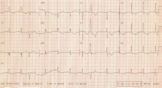
Electrocardiogram of an 86 year-old man with hypertension, coronary artery disease, possible previous myocardial infarction, left ventricular systolic dysfunction, admitted with recurrent chest and abdominal pains.
Rate 77/min
PR 0.18 sec
QRS 0.09 sec
QT 0.41 sec
QTc 0.47 sec
QRS axis –15º
Interpretation: Abnormal. Sinus rhythm, occasional premature ventricular complexes. Borderline left atrial abnormality. Minor intraventricular conduction delay. Diffuse ST-T abnormalities representing likely myocardial ischemia. Deep T wave inversion in precordial leads suggestive of proximal LAD stenosis.
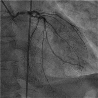
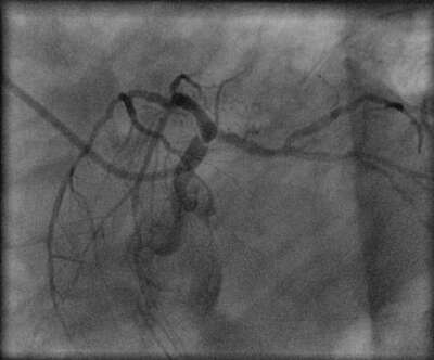
On the basis of elevated troponin I diagnosis of non-ST segment elevation myocardial infarction was made. The patient underwent cardiac catheterization and coronary angiogram which showed 90-95% left main coronary artery stenosis, 90% LAD, 95 % LCx, 90% marginal branch and 98 % dominant RCA stenosis with collaterals from the left system ( two representative views of left coronary artery are presented).
There was severe left ventricular systolic dysfunction.
Despite senility, significant left ventricular systolic dysfunction and mild renal failure the patient underwent successful revascularization with CABG.


<< back to ECG Decoder
|